
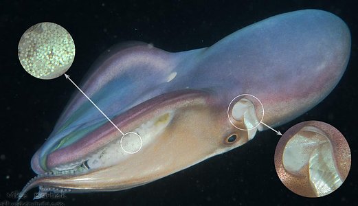
Figure. Tremoctopus gracilis, female brooding eggs held within the arm crown. Left insert - enlarged view of eggs with early embryos. Right insert - The curved antrior edge of the mantle opening suggests that the octopod was exhaling water through this opening while immediately below in the mantle opening a series of ridges suggest this is the primary site for inhaling. Photographed in situ while scuba diving at night in Janao Bay, Phillippines about two kilometers from land. © 2018 Mike Bartick
Below we present photographs of an egg mass taken from a female T. gracilis from Hawaiian waters. The octopod was captured in Hanauma Bay on Oahu, Hawaii in the summer of 2003. Liz Kumabe Maynard writes, "The animal was found floating in the Witches Brew area of HBay by visitors and eventually it was brought to shore where a combination of visitors and park rangers brought it to the attention of the HBay Education staff who then tucked it into the refrigerator .... It was quite a joint effort by all. My understanding was that it was just about dead when found with still some pulses of chromatophores." For comparison, drawings by Naef (1928) are provided from the Mediterranean T. violaceus.
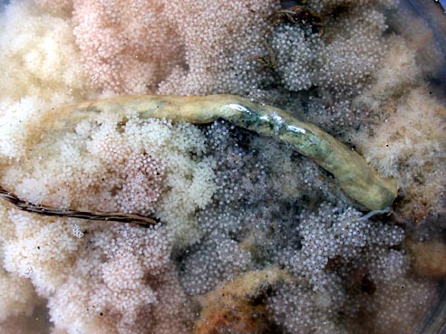
Figure. Dorsal view of a portion of the egg mass. The large worm-like structure in the center is the rod to which the egg mass is attached. The right end is broken off. The eggs in different regions of the egg mass exhibit different colors and structure. The white eggs to the left are in the earliest stage of development. The pink regions have embryos that are further developed and the dark grey embryos are still further along. On the right a rather fuzzy area above the rod is a region of mostly empty chorions where hatching has already occurred. Photograph by R. Young.
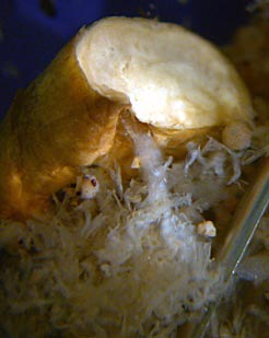
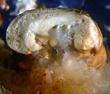
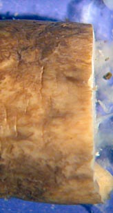
Figure. Various views of a piece of the rod (10 mm in longest cross-sectional width) that has been removed to show the structure of the rod and the way the egg strings attach. Left - Oblique view that shows several egg strings entering into depressions on the ventral surface of the rod. The largest has a white translucent appearance and looks like and inverted letter Y. This broad string is composed of many (hundreds?) of very thin strings. Center - Cross-sectional view that shows a cut through a major area of attachment. The ventral surface of the rod is very irregular with numerous holes where the translucent bundles of egg strings entering the rod. The rod itself appears to have a sponge-like structure with numerous small holes and channels. This porous structure, presumably, provides a large surface area for the attachment of the strings. The black structure in the center of this photograph is a sand grain that was picked up when the octopus was brought through the surf zone. Right - Dorsal view of the cut portion of the rod. The dorsal surface is smooth. Naef (1928) states that the rod or egg carrier looked almost like coral. The present rod does not resemble coral at all. Its composition is tough but flexible and appears to be entirely organic and not calcareous. A female is thought to carry two of these rods that are held by the dorsal arms (Naef, 1928).
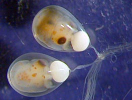
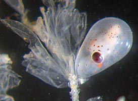
Figure. Left - Dorsal views of two embryos with white yolk sacs and with their egg stalks attaching to a portion of a larger, multistrand string. Right - Lateral view of an embryo lacking a yolk sac and ready to hatch. The egg has a short stalk that attaches to a heavier stalk. To the left of the embryo are empty chorions where hatching has already occurred. Photographs, using reflected light, by R. Young.
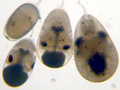
Figure. Various views of embryos in the four different stages found in the egg mass. Note the dark yolk sacs in the two middle embryos and the extension of the yolk into the embryo in the form of a dark Y-shaped structure. The embryo on the right is ready to hatch as indicated by the lack of an external yolk sac and the reverse orientation (the arms face the egg stalk). The embryo on the top-left measures 1.6 by 0.9 mm. Photographs, using transmitted light, by R. Young.
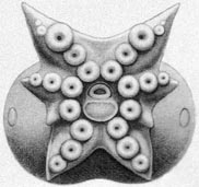
Figure. Oral view of the buccal crown of a late-stage embryo (Naef stage XIX) shows the arrangement of arms, suckers, central mouth and the adjacent (ventral) cut base of the external yolk sac. Drawing by Naef (1928).
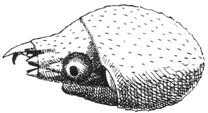
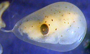
Figure. Lateral views of hatchling and near hatchling. Left - Hatchling, about 2.5 mm total length, from the Mediterranean Sea (Naef stage XX) with long dorsal arms I (dorsal) and short arm III (immediately in from of the eye). The lines over much of the hatchling represent Kölliker's organs. Note the extension of skin that forms a collar or cuff over the third arm and the bases of the other arms. Right - Embryo from Hawaii that is ready to hatch (Naef stage XX) with the thick collar over the arms and the relatively large dorsal arms. Kölliker's organs which appear as faint white marks are more obvious in the photograph below. The reddish-brown chromatophores are seen in this photograph and the ones below but are absent from Naef's drawings as they had faded after long preservation.

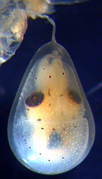
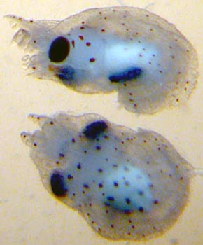
Figure. Left - Ventral view of the Naef stage XX hatchling from the Mediterranean. Drawing from Naef (1928). Middle - Ventral view of the Naef stage embro from Hawaii. Photograph by R. Young. Right - Side and dorsal views of two hatchlings from Hawaii that are slightly damaged. Photograph by R. Young.


