


New Subfamily
New Genus
Richard E. Young, Caroline Sanchez, and Valerie Allain


This tree diagram shows the relationships between several groups of organisms.
The root of the current tree connects the organisms featured in this tree to their containing group and the rest of the Tree of Life. The basal branching point in the tree represents the ancestor of the other groups in the tree. This ancestor diversified over time into several descendent subgroups, which are represented as internal nodes and terminal taxa to the right.
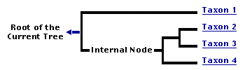
You can click on the root to travel down the Tree of Life all the way to the root of all Life, and you can click on the names of descendent subgroups to travel up the Tree of Life all the way to individual species.
For more information on ToL tree formatting, please see Interpreting the Tree or Classification. To learn more about phylogenetic trees, please visit our Phylogenetic Biology pages.
close boxIntroduction
Three specimens belonging to two distinct species, from the stomach of the fish Alepisaurus ferox, superficially look like sepiolids but lack several of the most basic features of that family and of the Sepioidea. We place these in a new subfamily but are uncertain how they fit into the phylogeny of the Sepiolidae or Sepiolida. A brief discussion of the relationships of the NewSubFamily is found on the Sepiolidae page.
Diagnosis
Sepiolid-like cephalopods:
- without lateral funnel adductor muscles.
- without secondary eyelids.
Characteristics
- Arms
- Arms without protective membranes or trabeculae.
- Large arm suckers globular in shape and with circularis muscle.
- Dorsal 6 arms connected by web; web deepest between arms III and IV.
Click on an image to view larger version & data in a new window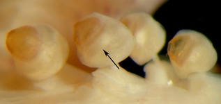
Figure. Venral view of suckers 3-6, arm III of New Species B, showing globular shape of suckers with small, smooth orifices. Arrow points to circularis muscle (shiny band). Photograph by R. Young.
- Tentacles
- Tentacular clubs small, with suckers only on distal part of club and in two series.
- Tentacular club keel restricted to non-sucker bearing region of club; keel curved inward.
- Protective membranes absent.
- Tentacular stalks long and very slender. Click on an image to view larger version & data in a new window
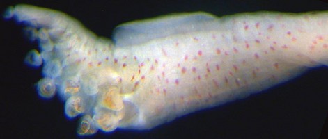
Figure. Oral view of the tentacular club of New Species B. Photograph by R. Young.
- Buccal connectives
- Attachments of ventral buccal connectives could not be determined.
- Head
- Eyes with corneas.
- Eyes without secondary eyelids.
Click on an image to view larger version & data in a new window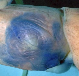
Figure. Side view of the left side of the head of New Species B showing the presence of a cornea and the absence of a secondary eyelid. Head stained with methylene blue stain. Photograph by R. Young.
- Funnel
- Funnel without lateral funnel adductor muscles.
- Funnel without funnel valve (uncertain in species A).
Click on an image to view larger version & data in a new window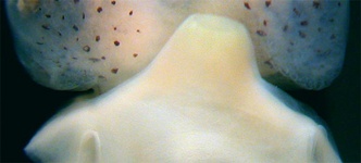
Figure. Ventral view of the funnel and posterior head of New Species A showing the absence of lateral funned adductor muscles. Photograph by R. Young.
- Mantle
- Stellate ganglia broadly separated; interstellate connective absent.
- Mantle component of the funnel locking-apparatus reaches anterior mantle margin.
- Anterior mantle margin does not protrude at points of locking-apparatuses.
Click on an image to view larger version & data in a new window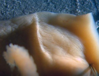
Figure. Frontal view of the mantle component of the funnel/mantle locking-aparatus of New Species B, showing that it extends, although barely, to the mantle margin. Photograph by R. Young.
- Fins
- Fins broadly separated posteriorly.
- Fins with anterior and posterior lobes.
Click on an image to view larger version & data in a new window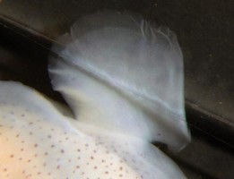
Figure. Ventral view of the right fin of New Species A showing anterior and posterior lobes. Part of the anterior lobe is folded under the fin. The edge of a glass slide is seen across the fin. Photograph by R. Young.
- Photophores
- Photophores absent.
- Gladius
- Gladius Y-shaped. Gladius extremely thin and delicate. Shell sac appears to be wider than gladius at anterior end.
Click on an image to view larger version & data in a new window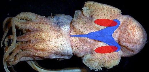
Figure. Dorsal view of New Species B with a drawing showing the approximate positions of the gladius (blue) and basal pockets (red). The basal pockets are entirely separate from the shell sac. Interpretation from a damaged gladius taken from a single specimen. Illustration by R. Young.
- Viscera
- Gills apparently without branchial canal*.
- Ventral mantle adductor muscle present but very reduced. Click on an image to view larger version & data in a new window
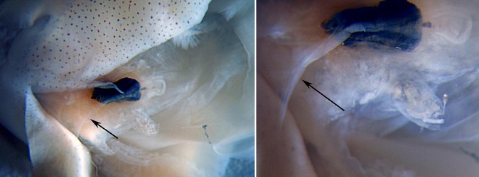
Figure. Ventral view of mantle cavity of New Species A. Arrows point to the ventral median adductor muscle located here at the leading edge of the ventral mantle septum and continuing onto the visceral mass passing to either side of the anus.
Comments
The gills were examined in the larger specimen and a branchial canal could not be detected. However, in such a small gill that is not in perfect condition, a branchial canal, if present would be very hard to detect. therefore, this character needs confirmation.
Distribution
Specimens taken from the stomach of Alepisaurus ferox captured within the EEZ of New Caledonia.
Title Illustrations
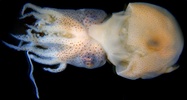
| Scientific Name | NewSpeciesA |
|---|---|
| Location | Off New Caledonia |
| Specimen Condition | Preserved |
| Sex | Male |
| Life Cycle Stage | Mature |
| View | Ventral |
| Size | 9.5 mm ML |
| Image Use |
 This media file is licensed under the Creative Commons Attribution-NonCommercial License - Version 3.0. This media file is licensed under the Creative Commons Attribution-NonCommercial License - Version 3.0.
|
| Copyright |
©

|
About This Page
University of Hawaii, Honolulu, HI, USA
Secretariat of the Pacific Community
Secretariat of the Pacific Community, Noumea, New Caledonia
Page copyright © 2019 , , and
 Page: Tree of Life
New Subfamily. New Genus.
Authored by
Richard E. Young, Caroline Sanchez, and Valerie Allain.
The TEXT of this page is licensed under the
Creative Commons Attribution-NonCommercial License - Version 3.0. Note that images and other media
featured on this page are each governed by their own license, and they may or may not be available
for reuse. Click on an image or a media link to access the media data window, which provides the
relevant licensing information. For the general terms and conditions of ToL material reuse and
redistribution, please see the Tree of Life Copyright
Policies.
Page: Tree of Life
New Subfamily. New Genus.
Authored by
Richard E. Young, Caroline Sanchez, and Valerie Allain.
The TEXT of this page is licensed under the
Creative Commons Attribution-NonCommercial License - Version 3.0. Note that images and other media
featured on this page are each governed by their own license, and they may or may not be available
for reuse. Click on an image or a media link to access the media data window, which provides the
relevant licensing information. For the general terms and conditions of ToL material reuse and
redistribution, please see the Tree of Life Copyright
Policies.
- First online 24 November 2006
- Content changed 24 November 2006
Citing this page:
Young, Richard E., Caroline Sanchez, and Valerie Allain. 2006. New Subfamily. New Genus. Version 24 November 2006 (under construction). http://tolweb.org/New_Genus/77759/2006.11.24 in The Tree of Life Web Project, http://tolweb.org/

