


Mixiomycetes
Mixia osmundae
Mixiales
Merje Toome and M. Catherine Aime
Introduction
Mixia osmundae is the only species currently known in Class Mixiomycetes. The fungus is an intracellular parasite of ferns in the genus Osmunda L. in which it causes small yellow to brown leaf spots. In cases of heavy infection, spots may coalesce to form larger lesions and even cover whole leaflets. Mixia is known only from Osmunda regalis L. in Japan and Taiwan, and O. cinnamomea L. in the United States (Kramer, 1958; Mix, 1947; Nishida, 1911; Sugiyama and Katumoto, 2008). However, analysis of available sequence data from public databases indicates that members may also be present in China and in France (Toome et al. 2013). Many aspects of the biology and ecology of this fungus are still unknown, since it is rarely found and not sufficiently studied.
Characteristics
When growing within a fern host, Mixia forms intercellular hyphal cells with many nuclei and few cross walls (termed septa), a condition known as coenocytic. Once the fungus reaches the surface of the host’s epidermal cells, it forms sac-like swellings which develop into non-septate, oblong to ellipsoid spore-producing (i.e., sporogenous) cells (24–60 × 9–2.5 µm). Tiny spores (3–4.5 × 1.5–2.5 µm) are formed on the surface of these sporogenous cells, creating a powdery layer on the lower side of fern leaves. As is true for most other aspects of the life cycle, for a long time it was debated whether these spores are meiospores or mitospores (i.e., sexually or asexually produced). However, recent genome sequencing analyses suggest that the spores are asexual and haploid (Toome et al. 2013).
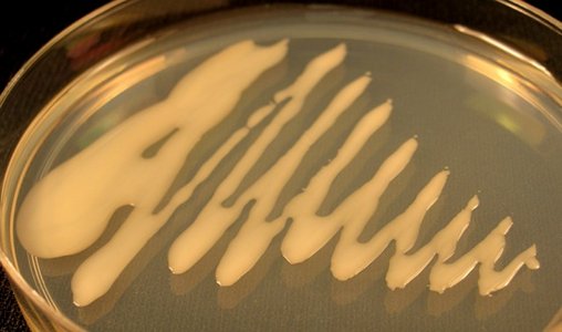
In culture, M. osmundae forms colonies composed of single yeast-like cells. The culture is white to cream colored at first with a slightly slimy texture that then turns pinkish with age. The cells in culture reproduce by budding; no hyphal growth has ever been detected on culture media. The production of coenocytic hyphae is a very rare condition in Basidiomycota and the sporogenous cells produced by Mixia are unique in the phylum.
Classification
Mixia osmundae was first described from Japan by Nishida (1911) as an ascomycete that he named Taphrina osmundae. Later, Mix (1947) described a second species, T. higginsii, as closely related to T. osmundae, but differing in host and by slight differences in sporogenous cell sizes. In 1958 Kramer created a new genus Mixia, into which he combined both species as Mixia osmundae (after determining that they were, indeed, the same species), and placed the genus in the Protomycetaceae (Taphrinales, Ascomycota). Mixia remained classified within phylum Ascomycota for more than eighty years, primarily due to superficial similarities between the sporogenous cells of Mixia and the asci produced by some Ascomycota. However, molecular and closer morphological studies of the sporogenous cells in the 1990’s provided multiple lines of evidence that Mixia belongs to Basidiomycota (Nishida et al., 1995), and that the structure previously interpreted as an ascus was instead a unique type of sporogenous cell (Bauer et al., 2006). Currently M. osmundae represents a monotypic taxon, Class Mixiomycetes in Pucciniomycotina (Aime et al., 2006; Bauer et al., 2006; Hibbett et al., 2007; Sjamsuridzal et al., 2002).
Genome
The genome of M. osmundae has been sequenced and annotated, which has allowed for a better understanding of some aspects of its biology (Toome et al. 2013). At 13.6 Mb M. osmundae has the smallest genome size sequenced to date for a plant pathogenic basidiomycete. The fungus is most likely a biotrophic plant pathogen because it has several enzyme sets to break down plant cell wall components but it does not have the enzymes to break the resulting molecules further down to sucrose. Therefore, as is common for other biotrophic fungi, M. osmundae needs to obtain its energy directly from the sucrose which is present only in living plants. A near complete set of mating and meiosis genes were detected in the genome of M. osmundae, however, all evidence seems to indicate that the observed spores and the yeast state produced in culture, are asexually produced and haploid, thus the sexual stage, if produced in nature, remains a mystery (Toome et al. 2013).Discussion of Phylogenetic Relationships
To date the phylogenetic position of Mixiomycetes has thoroughly been examined only with ribosomal DNA sequences, which support Mixiomycetes within Pucciniomycotina. Nevertheless, the sister group to Mixiomycetes has not yet been resolved (Aime et al., 2006). Phylogenomic data hint that Mixia is more closely related to Microbotryomycetes than Pucciniomycetes (Toome et al. 2013), however, more data are needed to better resolve the relationship between Mixiomycetes and the other classes of Pucciniomycotina.
References
Aime, M. C., P. B. Matheny, D. A. Henk, E. M. Frieders, R. H. Nilsson, M. Piepenbring, D. J. McLaughlin, L. J. Szabo, D. Begerow, J. P. Sampaio, R. Bauer, M. Weiß, F. Oberwinkler, and D. S. Hibbett. 2006. An overview of the higher-level classification of Pucciniomycotina based on combined analyses of nuclear large and small subunit rDNA sequences. Mycologia 98: 896905.
Bauer, R., D. Begerow, J. Sampaio, M. Weiβ, F. Oberwinkler. 2006. The simple-septate basidiomycetes: a synopsis. Mycological Progress 5: 4166.
Hibbett, D. S., M. Binder, J. F. Bischoff, M. Blackwell, P. F. Cannon, O. E. Eriksson, S. Huhndorf, T. James, P. M. Kirk, R. Lücking, T. Lumbsch, F. Lutzoni, P. B. Matheny, D. J. Mclaughlin, M. J. Powell, S. Redhead, C. L. Schoch, J. W. Spatafora, J. A. Stalpers, R. Vilgalys, M. C. Aime, A. Aptroot, R. Bauer, D. Begerow, G. L. Benny, L. A. Castlebury, P. W. Crous, Y.-C. Dai, W. Gams, D. M. Geiser, G. W. Griffith, C. Gueidan, D. L. Hawksworth, G. Hestmark, K. Hosaka, R. A. Humber, K. Hyde, J. E. Ironside, U. Kõljalg, C. P. Kurtzman, K.-H. Larsson, R. Lichtwardt, J. Longcore, J. Miądlikowska, A. Miller, J.-M. Moncalvo, S. Mozley-Standridge, F. Oberwinkler, E. Parmasto, V. Reeb, J. D. Rogers, C. Roux, L. Ryvarden, J. P. Sampaio, A. Schüßler, J. Sugiyama, R. G. Thorn, L. Tibell, W. A. Untereiner, C. Walker, Z. Wang, A. Weir, M. Weiß, M. M. White, K. Winka, Y.-J. Yao, and N. Zhang. 2007. A higher-level phylogenetic classification of the Fungi. Mycological Research 111: 509547.
Kramer, C.L. 1958. A new genus in the Protomycetaceae. Mycologia 50 (6): 916926.
Mix, A.J. 1947. Taphrina osmundae Nishida and Taphrina higginsii sp. nov. Mycologia 39 (1): 7176.
Nishida, H., K. Ando, Y. Ando, A. Hirata, and J. Sugiyama. 1995. Mixia osmundae: Transfer from the Ascomycota to the Basidiomycota based on evidence from molecules and morphology. Can. J. Bot., 73 (Suppl. 1): S660S666.
Sjamsuridzal, W., H. Nishida, A. Yokota. 2002. Phylogenetic position of Mixia osmundae inferred from 28S rDNA comparison. Journal of General and Applied Microbiology 48: 121123.
Sugiyama, J., K. Katumoto. 2008. Identity of the plasmodial slime mold Phytoceratiomyxa osmundae and the lectotypification of Taphrina osmundae, the basionym of Mixia osmundae. Mycoscience 49: 192198.
Toome, M., Ohm, R. A., Riley, R. W., James, T. Y., Lazarus, K. L., Henrissat, B., Albu, S., Boyd, A., Chow, J., Clum, A., Heller, G., Lipzen, A., Nolan, M., Sandor, L., Zvenigorodsky, N.,Grigoriev, I. V., Spatafora, J. W., Aime, M. C. 2013. Genome sequencing provides insight into the reproductive biology, nutritional mode and ploidy of the fern pathogen Mixia osmundae. New Phytologist, doi: 10.1111/nph.12653.
Title Illustrations
| Scientific Name | Mixia osmundae |
|---|---|
| Specimen Condition | Live Specimen |
| Image Use |
 This media file is licensed under the Creative Commons Attribution-NonCommercial License - Version 3.0. This media file is licensed under the Creative Commons Attribution-NonCommercial License - Version 3.0.
|
| Copyright |
©

|
About This Page
Development of this page was facilitated by the "Deep Hypha" Research Coordination Network and the "Assembling the Fungal Tree of Life" project (NSF award DEB-0732968).
Purdue University
M. Catherine Aime
Louisiana State University Agricultural Center
Correspondence regarding this page should be directed to Merje Toome at and M. Catherine Aime at
Page copyright © 2014 and M. Catherine Aime
 Page: Tree of Life
Mixiomycetes. Mixia osmundae. Mixiales.
Authored by
Merje Toome and M. Catherine Aime.
The TEXT of this page is licensed under the
Creative Commons Attribution-NonCommercial-ShareAlike License - Version 3.0. Note that images and other media
featured on this page are each governed by their own license, and they may or may not be available
for reuse. Click on an image or a media link to access the media data window, which provides the
relevant licensing information. For the general terms and conditions of ToL material reuse and
redistribution, please see the Tree of Life Copyright
Policies.
Page: Tree of Life
Mixiomycetes. Mixia osmundae. Mixiales.
Authored by
Merje Toome and M. Catherine Aime.
The TEXT of this page is licensed under the
Creative Commons Attribution-NonCommercial-ShareAlike License - Version 3.0. Note that images and other media
featured on this page are each governed by their own license, and they may or may not be available
for reuse. Click on an image or a media link to access the media data window, which provides the
relevant licensing information. For the general terms and conditions of ToL material reuse and
redistribution, please see the Tree of Life Copyright
Policies.
- First online 16 August 2005
- Content changed 21 January 2014
Citing this page:
Toome, Merje and M. Catherine Aime. 2014. Mixiomycetes. Mixia osmundae. Mixiales. Version 21 January 2014 (under construction). http://tolweb.org/Mixia_osmundae/51266/2014.01.21 in The Tree of Life Web Project, http://tolweb.org/


