


Microsporidia


References
Canning, E. U. 1998. Evolutionary relationships of Microsporidia. Pages 77-90 in Evolutionary Relationships among Protozoa (G. H. Coombs, K. Vickerman, M. A. Sleigh, and A. Warren, eds.) Chapman & Hall, London.
Fast, N. M., J. M. Logsdon, and W. F. Doolittle. 1999. Phylogenetic analysis of the TATA box binding protein (TBP) gene from Nosema locustae: evidence for a microsporidia-fungi relationship and spliceosomal intron loss. Molecular Biology and Evolution 16:1415-1419.
Fischer, W. M. and J. D. Palmer. 2005. Evidence from small-subunit ribosomal RNA sequences for a fungal origin of Microsporidia. Molecular Phylogenetics and Evolution 36:606-622.
Germot, A., H. Philippe, and H. Le Guyader. 1997. Evidence for loss of mitochondria in Microsporidia from a mitochondrial-type HSP70 in Nosema locustae. Molecular and Biochemical Parasitology 87:159-168.
Hirt, R. P., B. Healy, C. R. Vossbrinck, E. U. Canning, and T. M. Embley. 1997. A mitochondrial Hsp70 orthologue in Vairimorpha necatrix: Molecular evidence that microsporidia once contained mitochondria. Current Biology 7:995-998.
Hirt, R. P., J. M. Logsdon, Jr., B. Healy, M. W. Dorey, W. F. Doolittle, and T. M. Embley. 1999. Microsporidia are related to fungi: evidence from the largest subunit of RNA polymerase II and other proteins. Proceedings of the National Academy of Sciences (USA) 96:580-585.
Keeling, P. J., M. A. Luker, and J. D. Palmer. 2000. Evidence from beta-tubulin phylogeny that microsporidia evolved from within the fungi. Molecular Biology and Evolution 17:23-31.
Keeling, P. J. and G. I. McFadden. 1998. Origins of microsporidia. Trends Microbiol. 6:19-23.
Li, J., S. K. Katiyar, A. Hamelin, G. S. Visvesvara, and T. D. Edlind. 1996. Tubulin genes from AIDS-associated microsporidia and implications for phylogeny and benzimidazole sensitivity. Molecular and Biochemical Parasitology 78:289-295.
Lom, J. and F. Nilsen. 2003. Fish microsporidia: fine structural diversity and phylogeny. International Journal for Parasitology 33:107-127.
Sprague, V., J. J. Becnel, and E. I. Hazard. 1992. Taxonomy of phylum Microspora. Critical Reviews in Microbiology 18(5-6):285-395.
Van de Peer, Y., A. Ben Ali, and A. Meyer. 2000. Microsporidia: accumulating molecular evidence that a group of amitochondriate and suspectedly primitive eukaryotes are just curious fungi. Gene 246:1-8.
Vossbrinck, C. R., T. G. Andreadis, J. Vavra, and J. J. Becnel. 2004. Molecular phylogeny and evolution of mosquito parasitic microsporidia (Microsporidia : Amblyosporidae). Journal of Eukaryotic Microbiology 51(1):88-95.
Vossbrinck, C. R. and B. A. Debrunner-Vossbrinck. 2005. Molecular phylogeny of Microsporidia: ecological, ultrastructural and taxonomic considerations. Folio Parasitologica 52:131-142.
Title Illustrations
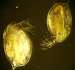
| Scientific Name | Binucleata daphniae infecting Daphnia magna |
|---|---|
| Comments | accumulation of microsporidian spores in the hypodermis makes the infected host conspicuously opaque (left individual) when compared with the non infected host (right individual) |
| Specimen Condition | Live Specimen |
| Life Cycle Stage | spores |
| Copyright |
© 2008

|
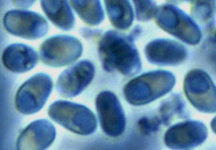
| Scientific Name | Binucleata daphniae |
|---|---|
| Comments | spores(about 5.0 x 2.5 μm in size) isolated from infected hypodermal cells of infected Daphnia. Each spore shows a vacuole-like area in the posterior pole of the spore. This is a specific character of microsporidian spores. |
| Specimen Condition | Live Specimen |
| Life Cycle Stage | spores |
| Copyright |
© 2008
|
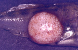
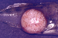
| Scientific Name | Glugea anomala |
|---|---|
| Comments | host cell of the stickleback Gasterosteus (Pisces:Teleostei) filled with microsporidian spores is hypertrophied to a macroscopic cyst (xenoma). |
| Specimen Condition | Live Specimen |
| Life Cycle Stage | spores |
| Copyright | © 2004 Ronny Larsson |

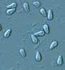
| Scientific Name | Glugea anomala |
|---|---|
| Comments | fresh spores isolated from the tissue cyst of the stickleback. Many fish microsporidia have a very large vacuole in their spores. |
| Specimen Condition | Live Specimen |
| Life Cycle Stage | spores |
| Copyright | © 2004 Ronny Larsson |
About This Page
Page copyright © 2012
All Rights Reserved.
- First online 09 January 2008
- Content changed 09 January 2008
Citing this page:
Tree of Life Web Project. 2008. Microsporidia. Version 09 January 2008 (temporary). http://tolweb.org/Microsporidia/2378/2008.01.09 in The Tree of Life Web Project, http://tolweb.org/


